
Pulp Exposure? How Modern Dentistry Preserves Natural Teeth with Vital Pulp Therapy2026-08-19
Pulp Exposure? How Modern Dentistry Preserves Natural Teeth with Vital Pulp Therapy
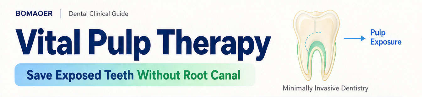
Deep caries, trauma, or accidental exposure during cavity prep—when does vital pulp therapy work, and when is it too late? This clinical guide walks you through indications, techniques, and material selection for predictable outcomes.
Introduction
When pulp exposure occurs during dental treatment, clinicians face a classic fork in the road: remove the entire pulp and proceed with root canal therapy, or attempt to preserve whatever remains viable?
For decades, root canal treatment was the default answer. But advances in biomaterials and a growing emphasis on minimally invasive dentistry have shifted the landscape. Vital pulp therapy now offers a middle ground—not wholesale removal, but selective preservation, allowing the tooth to retain its vitality even after restorative work.
This article lays out a clinical decision-making framework for vital pulp therapy: when to consider it, which technique fits which scenario, and how to execute each step with precision.
1. What Exactly Is Vital Pulp Therapy?
At its core, vital pulp therapy operates on a simple premise: when pulp tissue still retains its self-repair capacity, the clinician's role is to remove irritants, control inflammation, seal the wound, and let the pulp heal on its own.
Unlike root canal therapy, which follows a "clean out everything" philosophy, vital pulp therapy takes a "minimal necessary intervention" approach. It acknowledges that not all exposed pulp is doomed—some can recover if given the right biological environment.
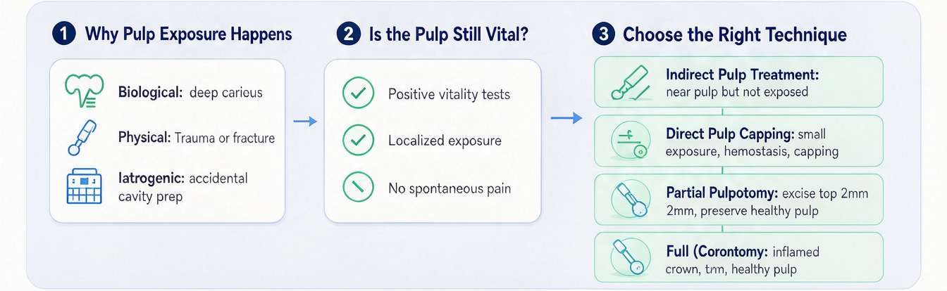
Clinically, a tooth is a good candidate for pulp therapy when it meets three basic criteria:
- Pulp vitality tests (cold, heat, or EPT) return a positive response, indicating functioning neurovascular supply;
- Exposure is localized, with no signs of inflammation spreading into the radicular pulp;
- The patient reports no spontaneous pain or tenderness to percussion, which would suggest irreversible pulpitis.
2. Common Culprits Behind Pulp Exposure
Pulp exposure in clinical practice typically stems from one of three pathways:
| Category | Examples |
|---|---|
| Biological | Deep carious lesions breaking through the dentinal barrier |
| Physical | Trauma from falls, sports injuries, or fractures exposing the pulp chamber |
| Iatrogenic | Accidental mechanical exposure during caries excavation or crown preparation, especially in deep lesions |
One important clinical nuance: exposed pulp doesn't always hurt right away. Some patients present with only transient sensitivity to cold or mild discomfort on chewing. That's why a thorough diagnostic workup—not just symptom history—is essential before making treatment decisions.
3. Four Techniques, One Goal: Know Which to Choose
Indirect Pulp Treatment – For Cases Where the Pulp Hasn't Been Breached
Deep decay has come close to the pulp but the dentinal layer remains intact. Instead of excavating all the way in, the clinician removes the bulk of infected dentin, leaves a thin affected layer over the pulp, places a biocompatible liner, and seals the tooth definitively. The remaining dentin acts as a natural barrier, and with the help of the liner, the pulp forms reparative dentin over time.
Direct Pulp Capping – For Pinpoint Exposures
A small exposure—typically 1mm or less—occurs during excavation or trauma. After achieving hemostasis with sterile saline, the clinician places a bioactive capping material directly onto the exposed pulp surface, then seals the site with a permanent restoration. The key prerequisite here is minimal bleeding and no purulent exudate.
Partial Pulpotomy – For Superficial Inflammation
When the exposure is slightly larger or the superficial layer of pulp shows signs of irreversible change, but deeper tissue remains healthy, the clinician uses a sterile bur to excise the top 2mm of inflamed pulp tissue. The remaining healthy pulp is then capped. This approach is particularly common in young permanent teeth following traumatic injury.
Full (Coronal) Pulpotomy – For Inflammation Confined to the Crown
If inflammation involves the entire coronal pulp but has not yet reached the radicular pulp (within the root canals), the entire coronal portion is removed while the radicular pulp is preserved. The remaining pulp continues to provide nutrition and sensory function to the tooth.
Choosing the right level is essentially a balancing act—removing just enough inflamed tissue to ensure healing, while preserving as much healthy tissue as possible. The decision hinges on accurate preoperative diagnosis and intraoperative visual assessment.
4. Step-by-Step: Six Critical Phases
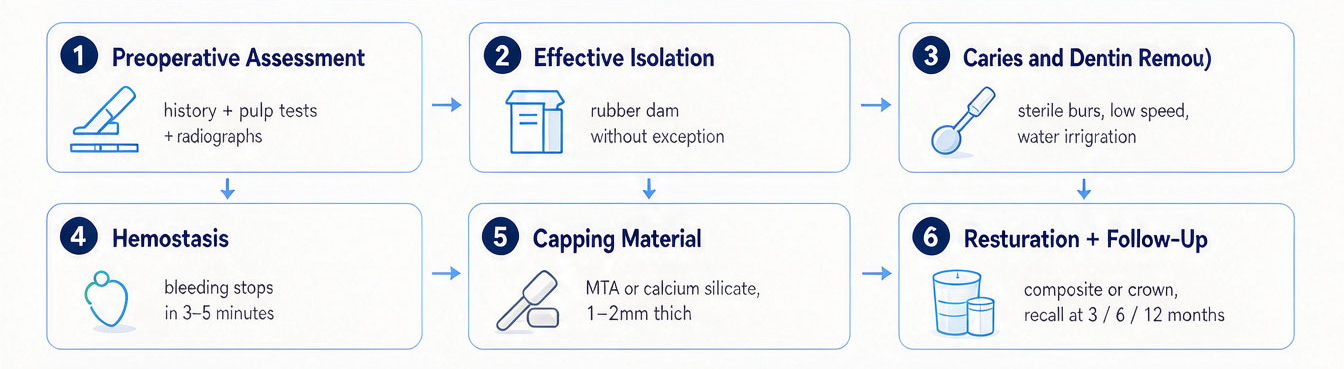
Success in vital pulp therapy is highly technique-sensitive. A misstep at any stage can tip a salvageable pulp toward irreversible damage. Here's a standardized workflow:
Phase 1 – Preoperative Assessment
Combine patient history, thermal/electrical pulp testing, and periapical radiographs to rule out irreversible pulpitis and apical pathology. This is the "gatekeeper" step—if the pulp is beyond saving, don't proceed.
Phase 2 – Effective Isolation
Place a rubber dam without exception. Salivary contamination of the exposed pulp surface is a guaranteed path to failure.
Phase 3 – Caries and Dentin Removal
Under magnification (loupes or microscope), remove infected dentin using sterile burs at low speed with copious water irrigation to prevent thermal injury to the pulp.
Phase 4 – Hemostasis and Wound Preparation
Gently irrigate the exposed site with sterile saline or a mild sodium hypochlorite solution. Apply gentle pressure with a sterile cotton pellet. The target endpoint: bleeding stops within 3–5 minutes, leaving a clean, dry surface.
Phase 5 – Placement of the Capping Material
Apply a bioceramic material (e.g., MTA or a calcium silicate–based cement) directly over the pulp wound, approximately 1–2mm thick. These materials do more than physically seal—they release calcium and silicate ions that actively promote dentin bridge formation.
Phase 6 – Definitive Restoration and Follow-Up
After the capping material sets, place a bonded restoration (composite resin or full crown) that provides a bacteria-tight seal. Schedule recalls at 3, 6, and 12 months for radiographic and vitality checks.
5. Material Matters: What Works and Why
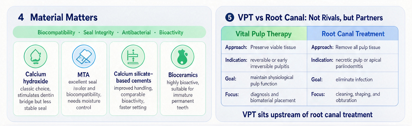
The material placed directly on the pulp is arguably the single most important clinical variable. It must satisfy four requirements simultaneously: biocompatibility, seal integrity, antibacterial properties, and bioactivity (the ability to stimulate regeneration).
| Material Class | Key Characteristics |
|---|---|
| Calcium hydroxide | Classic choice; stimulates dentin bridge formation but lacks long-term sealing stability |
| MTA | Gold standard for many clinicians; excellent seal and biocompatibility; handling requires moisture control |
| Calcium silicate–based cements | Improved handling over MTA; comparable bioactivity and sealing; faster setting options |
| Bioceramics | Newer generation; highly bioactive; particularly suitable for immature permanent teeth |
A crucial point often overlooked: the material doesn't "heal" the pulp on its own. It creates the environment—a protected, bacteria-free space—in which the pulp's own regenerative capacity can do the actual work.
6. Vital Pulp Therapy vs. Root Canal: Not Rivals, but Partners
It's a common misconception that vital pulp therapy is meant to "replace" root canal treatment. A more useful framing: they serve different stages of the same disease spectrum.
| Vital Pulp Therapy | Root Canal Treatment | |
|---|---|---|
| Approach to pulp | Preserve viable tissue | Remove all pulp tissue |
| Indication | Reversible pulpitis; early irreversible cases | Necrotic pulp; apical periodontitis |
| Goal | Maintain physiological pulp function | Eliminate infection; retain the tooth as a non-vital structure |
| Technical focus | Diagnosis and biomaterial placement | Cleaning, shaping, and obturation of canals |
These two approaches are not in opposition. Vital pulp therapy sits upstream of root canal treatment—when the disease crosses the line into irreversible territory, root canal therapy remains the dependable fallback.
7. When Does Vital Pulp Therapy Make the Most Sense?
Based on current evidence and clinical guidelines, the following scenarios offer the best risk–benefit profile for pulp therapy:
- Immature permanent teeth – Open apices and rich blood supply enhance healing capacity;
- Traumatic exposures – The pulp is mechanically exposed but not infected, giving it a clean starting point;
- Iatrogenic exposures ≤1mm – Early detection and minimal contamination improve outcomes;
- Deep carious exposures with controllable bleeding – No spontaneous pain history; inflammation appears localized.
For adult patients with deep carious exposures, more caution is warranted. Pulpal blood supply and regenerative capacity decline with age, so the success threshold shifts—but age alone is not an absolute contraindication. The decisive factor is the extent of inflammation, not the patient's chronological age.
Conclusion
The growing adoption of vital pulp therapy reflects a broader evolution in dentistry: from a purely ablative mindset to one that prioritizes biological preservation. This shift has been driven by better materials, finer diagnostic tools, and a deeper understanding of pulp biology.
That said, pulp therapy isn't a magic bullet. It demands sharper diagnostic acumen and stricter procedural discipline from the clinician—not to mention patient compliance with follow-up visits. But when it succeeds, the payoff is significant: a tooth that retains its natural proprioception, occlusal sensation, and structural integrity.
For the clinician, integrating vital pulp therapy into practice isn't about abandoning root canal treatment—it's about expanding the toolbox and making more nuanced decisions at the chairside.
FAQs
Q1: What is the long-term success rate of vital pulp therapy?
In properly selected cases using bioceramic materials, direct pulp capping shows 5-year success rates of 85–90%. However, individual outcomes vary, and regular follow-up is non-negotiable.
Q2: Is vital pulp therapy less effective in adults than in children?
Children and adolescents have richer pulpal blood supply and greater regenerative potential. But adults with small exposures and minimal inflammation can also achieve excellent outcomes—age alone shouldn't disqualify a case.
Q3: Does the pulp fully recover after treatment?
In most successful cases, the pulp forms a new dentin bridge to seal the wound and gradually restores normal vascular and neural function. Long-term monitoring remains important.
Q4: Is the procedure painful for the patient?
The procedure is performed under local anesthesia, so patients experience no pain during treatment. Postoperative sensitivity is usually mild and self-limiting.
Q5: If vital pulp therapy fails, can the tooth still have root canal treatment later?
Absolutely. Pulp therapy doesn't compromise the tooth's suitability for root canal treatment if needed later—the two are sequential, not mutually exclusive.
About BOMAOER
Henan Bomaoer Commercial Cooperation specializes in supplying high-value dental equipment and consumables to clinical practices worldwide. Our product range covers everything you need for vital pulp therapy workflows—from high-speed handpieces and surgical burs to curing lights and rubber dam kits. We believe that quality materials should be accessible, so clinicians can focus on what matters most: patient care.
Website: www.bomaoer.com
WhatsApp / Tel: +86 13781942705
Email: bomaoer@hotmail.com

- Company Info
- Feedback
- Customer Reviews
- About Us
- Contact Us
- News
- Privacy Policy
- User Center
- Forget Password
- My Orders
- Tracking Order
- My Account
- Wanting to cooperate with us?
- Register
- Not Satisfied with our products or service?
- Payment & Shipping
- Customs & Taxes
- Locations We Ship To
- Shipping Methods
- Payment Methods
- Newsletter



 Leave a message